What is the function of chloroplasts and where are they found?
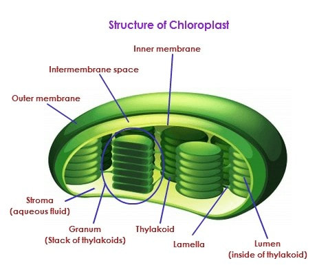
The chloroplasts are double membrane bound organelles found in higher plants and are the site of photosynthesis. The outer and the inner membrane of the chloroplast enclose a semi-gel-like fluid known as the stroma. Stroma is a alkaline, aqueous fluid which is protein rich and is present within the inner membrane of the chloroplast. The thylakoid system is suspended in the stroma. The thylakoid system is a collection of membranous sacks called thylakoids. The chlorophyll is found in the thylakoids.
Function of chloroplasts:
These organelles conduct photosynthesis. They absorb sunlight and convert it into sugar molecules and also produce free energy stored in the form of ATP and NADPH through photosynthesis.
Draw the diagram of a chloroplast
Describe the structure and function of a vacuole.
Vacuoles are membrane-bound sacs within the cytoplasm of a cell that function in several different ways. In mature plant cells, vacuoles tend to be very large and are extremely important in providing structural support, as well as serving functions such as storage, waste disposal, protection, and growth. Vacuoles in animal cells, however, tend to be much smaller, and are more commonly used to temporarily store materials or to transport substances. They also help in the intracellular digestion and disposal of cellular waste.
What is an endoplasmic reticulum and what is its function?
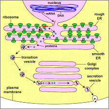
The Endoplasmic Reticulum is an extensive network of flattened sacs and tubes. Tubules and sacs of the ER enclose an interior space that is separate from the cytoplasmic fluid. It is of two types:
RER – Its surface is studded with protein manufacturing units (ribosomes) that gives it the rough appearance. A ribosome binds ER only when it begins to synthesize protein for secretion.
SER – They have no ribosomes. It is connected to the nuclear envelope. It functions are:
What is the golgi body and what is its function?
The Golgi apparatus is an organelle present in most eukaryotic cells. It is made up of membrane-bound sacs, and is also called a Golgi body, Golgi complex, or dictyosome.
The job of the Golgi apparatus is to process and bundle macromolecules like proteins and lipids as they are synthesized within the cell. The Golgi apparatus is sometimes compared to a post office inside the cell since one major function is to modify, sort, and package proteins to be secreted.
This organelle is also important in other ways, specifically in the transport of lipids throughout the cell and the creation of lysosomes.
Give a diagram to show how endoplasmic reticulum and golgi complex work together.
Vacuoles are membrane-bound sacs within the cytoplasm of a cell that function in several different ways. In mature plant cells, vacuoles tend to be very large and are extremely important in providing structural support, as well as serving functions such as storage, waste disposal, protection, and growth. Vacuoles in animal cells, however, tend to be much smaller, and are more commonly used to temporarily store materials or to transport substances. They also help in the intracellular digestion and disposal of cellular waste.
What is an endoplasmic reticulum and what is its function?
The Endoplasmic Reticulum is an extensive network of flattened sacs and tubes. Tubules and sacs of the ER enclose an interior space that is separate from the cytoplasmic fluid. It is of two types:
RER – Its surface is studded with protein manufacturing units (ribosomes) that gives it the rough appearance. A ribosome binds ER only when it begins to synthesize protein for secretion.
SER – They have no ribosomes. It is connected to the nuclear envelope. It functions are:
- Synthesis of steroids and lipids.
- Metabolism of carbohydrates.
- Regulation of calcium levels.
- Drug detoxification
What is the golgi body and what is its function?
The Golgi apparatus is an organelle present in most eukaryotic cells. It is made up of membrane-bound sacs, and is also called a Golgi body, Golgi complex, or dictyosome.
The job of the Golgi apparatus is to process and bundle macromolecules like proteins and lipids as they are synthesized within the cell. The Golgi apparatus is sometimes compared to a post office inside the cell since one major function is to modify, sort, and package proteins to be secreted.
This organelle is also important in other ways, specifically in the transport of lipids throughout the cell and the creation of lysosomes.
Give a diagram to show how endoplasmic reticulum and golgi complex work together.
Describe the structure and function of ribosomes.
Ribosomes are protein manufacturing units that are made up of RNA and proteins. Ribosomes can be found floating within the cytoplasm or attached to the endoplasmic reticulum. Ribosomes can prepare proteins from all amino acids. The ribosomes are made up of two subunits - a small and a large subunit. They assemble amino acids to form specific proteins, proteins are essential to carry out cellular activities. The proteins that are synthesized by the ribosomes present in the cytoplasm are used in the cytoplasm itself. The proteins produced by the bound ribosomes are transported outside the cell.
Describe the structure and function of mitochondria.
Mitochondria are known as the powerhouses of the cell. Mitochondria are small organelles floating free throughout the cell. Mitochondria are made of two membranes. The outer membrane covers the organelle and contains it like a skin. The inner membrane folds over many times and creates layered structures called cristae. The fluid contained in the mitochondria is called the matrix. The folding of the inner membrane increases the surface area inside the organelle. Since many of the chemical reactions happen on the inner membrane, the increased surface area creates more space for reactions to occur. The matrix is filled with water and proteins (enzymes).
Describe the structure and function of lysosome.
Lysosomes are single –membrane bound structures that contain digestive enzymes. They engulf the bacteria and other cellular debris of animal cells. They are also known as cells suicidal bags. They contain hydrolytic enzymes.
Describe the structure and function of centrioles.
Centrioles are a pair of rod shaped structures that lie perpendicular to each other and are located just outside the nucleus. They are made up of microtubules.
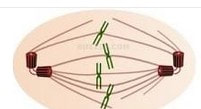
The centrioles play a major role in cell division. The centrioles help in the formation of the spindle fibers that separate the chromosomes during cell division (mitosis).
Ribosomes are protein manufacturing units that are made up of RNA and proteins. Ribosomes can be found floating within the cytoplasm or attached to the endoplasmic reticulum. Ribosomes can prepare proteins from all amino acids. The ribosomes are made up of two subunits - a small and a large subunit. They assemble amino acids to form specific proteins, proteins are essential to carry out cellular activities. The proteins that are synthesized by the ribosomes present in the cytoplasm are used in the cytoplasm itself. The proteins produced by the bound ribosomes are transported outside the cell.
Describe the structure and function of mitochondria.
Mitochondria are known as the powerhouses of the cell. Mitochondria are small organelles floating free throughout the cell. Mitochondria are made of two membranes. The outer membrane covers the organelle and contains it like a skin. The inner membrane folds over many times and creates layered structures called cristae. The fluid contained in the mitochondria is called the matrix. The folding of the inner membrane increases the surface area inside the organelle. Since many of the chemical reactions happen on the inner membrane, the increased surface area creates more space for reactions to occur. The matrix is filled with water and proteins (enzymes).
Describe the structure and function of lysosome.
Lysosomes are single –membrane bound structures that contain digestive enzymes. They engulf the bacteria and other cellular debris of animal cells. They are also known as cells suicidal bags. They contain hydrolytic enzymes.
Describe the structure and function of centrioles.
Centrioles are a pair of rod shaped structures that lie perpendicular to each other and are located just outside the nucleus. They are made up of microtubules.
The centrioles play a major role in cell division. The centrioles help in the formation of the spindle fibers that separate the chromosomes during cell division (mitosis).
What are the distinguishing characters of the structure of the nucleus?
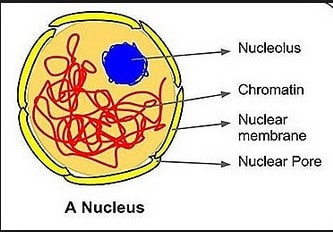
The cell nucleus is a membrane bound structure that contains the cell's hereditary information and controls the cell's growth and reproduction. It is the command center of a eukaryotic cell and is commonly the most prominent organelle in a cell. The cell nucleus is bound by a double membrane called the nuclear envelope. This membrane separates the contents of the nucleus from the cytoplasm.
The nucleus is the organelle which houses chromosomes. Chromosomes consist of DNA, which contains heredity information and instructions for cell growth, development, and reproduction.
Nucleoplasm is the gelatinous substance within the nuclear envelope. It is composed mainly of water with dissolved salts, enzymes, and organic molecules suspended within.
Nucleolus is a specialized region within the nucleus. It a membrane less structure composed of RNA and proteins. The nucleolus helps to synthesize ribosomes.
Draw the structure of the nucleus and label its parts.
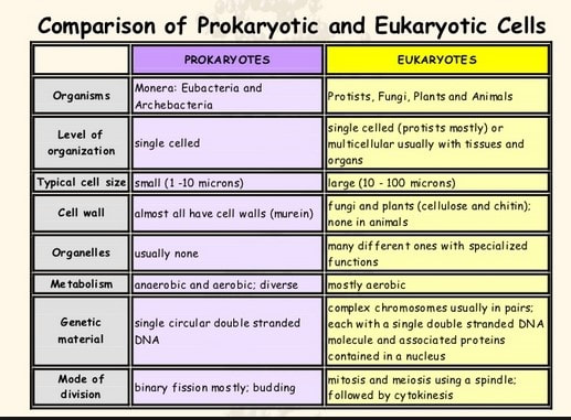
A comparison between prokaryotic and eukaryotic cells
What is microscopy?
Microscopy is the technical field of using microscope to view samples and objects that cannot be seen with naked eyes. Microscopy helps in analysis of tissues, cells or organelle preparation.
How many types of microscopes are available?
Microscopes can be basically classified into two types - Simple and compound microscope
Compound microscopes are of two types - Optical microscope and electron microscope.
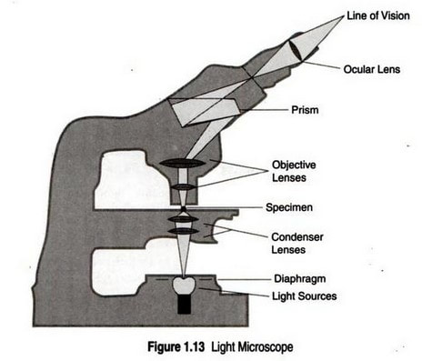
What is an optical microscope?
An optical light microscope uses optical lens and light wave for magnification. Mostly all microscopes have two lenses. The lens system nearest to the eye is called eye piece and that nearest to the specimen is called objective. An optical microscope magnifies an image by about 1000 to 1500 times.
What are the limitations of light microscope?
Microscopy is the technical field of using microscope to view samples and objects that cannot be seen with naked eyes. Microscopy helps in analysis of tissues, cells or organelle preparation.
How many types of microscopes are available?
Microscopes can be basically classified into two types - Simple and compound microscope
- Simple microscope - It works like a biconvex lens and uses a single lens
- Compound microscope – It has two separate lens systems for greater magnification
Compound microscopes are of two types - Optical microscope and electron microscope.
What is an optical microscope?
An optical light microscope uses optical lens and light wave for magnification. Mostly all microscopes have two lenses. The lens system nearest to the eye is called eye piece and that nearest to the specimen is called objective. An optical microscope magnifies an image by about 1000 to 1500 times.
What are the limitations of light microscope?
- Only images which are dark and can be viewed effectively.
- Live cells in particular generally lack sufficient contrast to be studied successfully. Internal structures are colourless and transparent so they need to be stained using selective dyes. In such case cells need to be killed and fixed.
What is an electron microscope?
Fundamental principles of an electron microscope and light microscope are same except that an electron microscope uses electromagnetic lenses and to focus a high velocity electron beam instead of visible light. A very high resolution can be achieved by using electron beam with far small wavelength. It magnifies the object by 10000 to 50000 times.
Fundamental principles of an electron microscope and light microscope are same except that an electron microscope uses electromagnetic lenses and to focus a high velocity electron beam instead of visible light. A very high resolution can be achieved by using electron beam with far small wavelength. It magnifies the object by 10000 to 50000 times.
 RSS Feed
